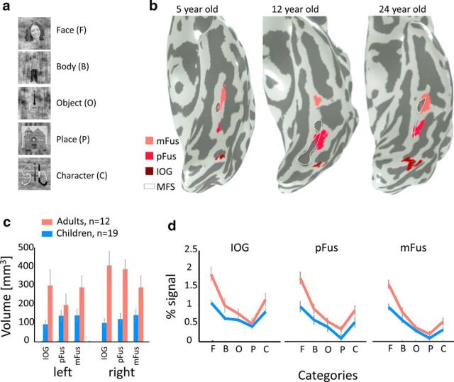
Figure 2.
Development of face-selective regions in the ventral stream. a, Example face, body, object, place, and character stimuli used in the localizer experiment. b, Face-selective regions in the ventral occipitotemporal cortex in the left hemisphere of three example subjects. Face-selective regions were defined using the contrast faces > objects, places, words, bodies (t > 3, voxel level), in the inferior occipital gyrus (IOG faces, dark red), in the posterior fusiform (pFus faces, red), and mid-fusiform (mFus faces, pink). White contour, mid fusiform sulcus (MFS). c, The average volume of face-selective regions (IOG, pFus, and mFus) in left and right hemispheres of 19 children (blue) and 12 adults (orange). Adult face-selective regions are significantly larger than those of children (F(1,157) = 62.91, p < 0.001). Error bars, standard error of mean (SEM) across participants of an age group. d, Independent analysis of percentage signal change in the three face-selective regions across five stimulus categories depicted in a. Result reveals a significant age-by-category interaction (F(4,410) = 8.43, p < 0.001) and significantly higher responses for faces in adults than in children (F(1,82) = 55.61, p < 0.001). Error bars, SEM across participants of an age group. F, Face; B, body; O, object; P, place; C, character.

