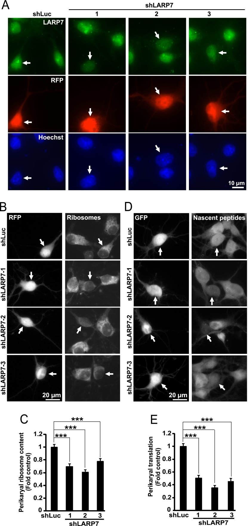
Fig. 7.
Reduced neuronal ribosome content and inhibition of protein synthesis after knockdown of LARP7 in hippocampal neurons. DIV4 (A) or DIV6 (B-E) rat hippocampal neurons were cotransfected with expression vectors for RFP and shRNAs against LARP7 or a control shRNA (shLuc, 0.2 + 0.6 μg of plasmid DNAs/2*105 neurons, respectively); in d-E, 0.1 μg EGFP expression vector was used instead of RFP. A, Representative images of transfected neurons (i.e. RFP-positive) that were fixed and immunostained for endogenous LARP7 at 72h post-transfection (arrows). Note that shLARP7 constructs reduced expression of endogenous LARP7 including disruption of the LARP7 signal in the nucleoli. B–C, After 3 days, perikaryal ribosome content was evaluated in RFP-positive cells by measuring intensity of ribosomal staining with NeuroTrace Green. For quantification, the ribosomal signal was normalized against DNA content in the nucleus as determined by counterstaining with Hoechst-33258. B, Representative images of transfected (i.e. RFP-positive) neurons whose ribosomes were stained with NeuroTrace Green (arrows). C, Quantification of ribosomal content in transfected neurons. shRNAs reduced mean ribosomal content. Data represent means ±S.E. of at least 72 cells from three independent experiments; ***, p < 0.001. D–E, Three days after transfection, O-propargyl-puromycin (OPP) was added to the culture medium to label nascent peptides which were visualized after fixation by fluorescent Click iT chemistry that was followed by GFP immunostaining. D, Representative images of transfected (i.e. GFP-positive) neurons with nascent peptide accumulation (arrows). E, Nascent peptide accumulation in the perikarya was reduced by shLARP7 constructs suggesting global translation deficiency. Data represent means ±S.E. of at least 54 individual cells from two independent experiments; ***, p < 0.001. Signal specificity controls for NeuroTrace staining of ribosomes (loss of staining after RNase A treatment) and OPP labeling of nascent peptides (loss of signal in cycloheximide-treated neurons) are presented in supplemental Fig. S5.

