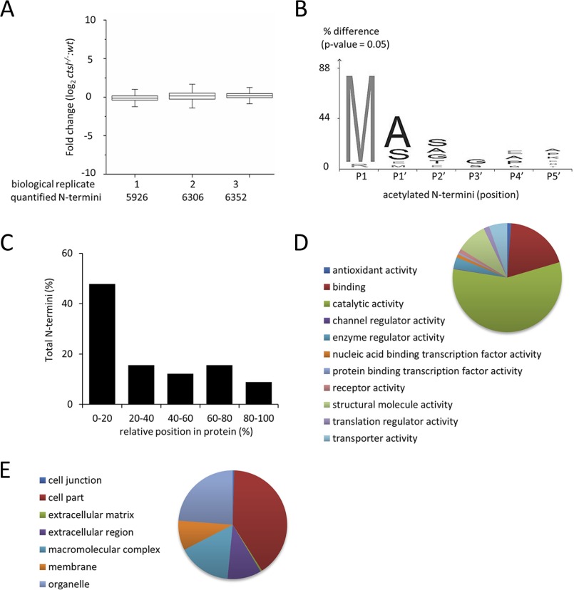
Fig. 6.
Identification of N-terminal peptides from FFPE liver tissues of C57BL/6 wild-type mice and C57BL/6 cathepsin L (Ctsl−/−) knock out mice. (A) Fold change distribution and Shapiro–Wilk normalization test (p value) of acetylated N termini and chemically dimethylated (naturally unmodified) N termini from three biological replicates (n = 3). (B) Visualization of N-α acetylation pattern in cathepsin L deficient tissue. Sequence logo was generated using iceLogo (60). (C) Positional clustering of acetylated and chemically dimethylated N termini from three biological replicates (n = 3). Gene Ontology database analysis of (D) molecular function and (E) cellular components of N-terminal peptides consistently identified all biological replicates (n = 3).

