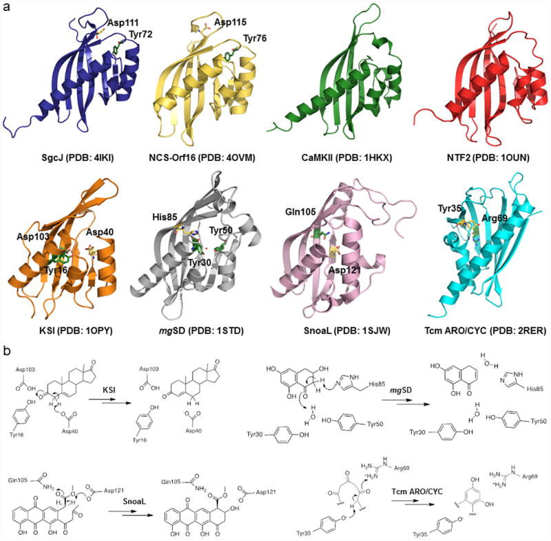
Figure 4.

Structure comparison between SgcJ and selected homologous in the NTF2-like superfamily. (a) Each structure shows a curved antiparallel β-sheet wall with a group of α-helices on one side of the wall to form the cone-like shapes. The putative general base and acid are shown as yellow and green sticks, respectively. The water molecule in the structure of mgSD is depicted by a red dot. Given in parentheses are PDB accession codes for each of the structures. (b) The proposed mechanisms for KSI, mgSD, SnoaL, and Tcm ARO/CYC, featuring the conserved general acid-base pairs to catalyze the initial steps of reactions.
