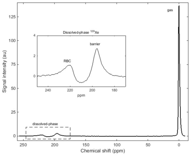
Fig. 2.
129Xe MR spectrum from a healthy 16-year-old girl, subject HV-17. Three spectral peaks are observed: the bulk gas-phase peak (0 ppm) and two dissolved-phase peaks (enlarged in the inset). The other two peaks originate from hyperpolarized 129Xe dissolved in the red blood cells and the barrier tissues (blood plasma and lung parenchymal tissue) peaks at ~212 ppm and ~197 ppm, respectively. ppm parts per minute

