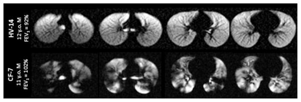
Fig. 4.
129Xe ventilation images for a healthy 12-year-old boy (HV-14, top row) and an 11-year-old boy with cystic fibrosis (CF-7, bottom row). The homogeneous signal seen throughout the lungs of the healthy control indicates that all regions of the lung are well ventilated. In contrast, the hyperpolarized 129Xe images of the child with cystic fibrosis display numerous regions of low signal intensity, indicating that ventilation is substantially impaired in multiple regions of the lungs

