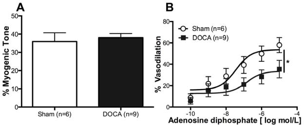
Figure 1. DOCA-salt treatment impairs endothelium dependent dilation in the MCA.
Endothelium functions in the MCAs were measured using ADP. There was no difference in percent myogenic tone between the two groups (A). Percent change in dilation was reduced in DOCA-salt MCAs (B). *p <0.05, Sham vs DOCA, two-way ANOVA. Values are mean± SEM. MCAs were mounted in a pressure myograph and kept in warm calcium PSS.

