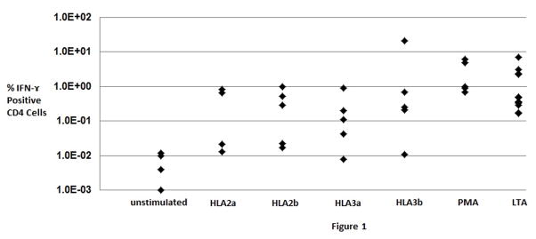
Fig. 1.
Response of healthy subject peripheral blood mononuclear cells to stimulation by HLA-A02, HLA-A03, or virus large T antigen (LTA) peptides as measured by flow cytometric assessment of % CD4 positive T-cells producing interferon-γ. CD8 positive T-cells reactive to viral and HLA peptides could not be consistently detected with the cell numbers analyzed. The polymorphic region of each HLA allele studied was represented by two peptides (sub-labeled a and b). Phorbol myristate acetate (PMA) was used as a positive control for stimulation, while unstimulated cells were used as a negative control to assess the background signal in the assay. Note the log scale on the Y-axis indicating considerable subject-to-subject differences in response. Each diamond represents a separate healthy subject (n= 4–5 for most conditions, n=12 for LTA peptides).

