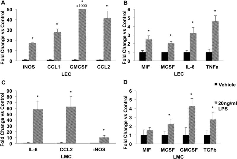
Figure 3.

LPS causes activation of proinflammatory cytokines and chemokines in LECs and LMCs. LECs and LMCs were treated either with PBS (vehicle control) or 20ng/ml LPS for 24 hours. Gene expression was analyzed using the ΔΔCt method as described in the Materials and Methods. A) LECs demonstrated a dramatic increase in the expression of iNOS, CCL1, CCL2, and GMCSF (over 1000 fold) in the LPS-treated group as compared to control group. B) LPS treated LECs also significantly increased expression of MIF, MCSF, IL-6, and TNFα. C) IL-6, CCL2, and iNOS expression was highly up regulated in response to LPS stimulation in LMCs. D) LPS treated LMCs also showed a significant increase in the expression of MIF, MCSF, GMCSF, and TGFβ as compared to the vehicle treated cells. Data are presented as Mean ± SEM. N=3; * denotes significance at p<0.05.
