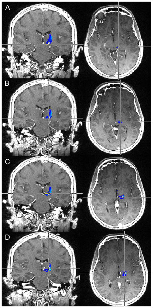
Figure 4.
Preoperative MRI-postoperative CT fusion for Patient 5. Image fusion demonstrates excellent placement of the DBS electrode within the VPL/VPM thalamus. Contacts 1, 2, and 3 (B, C, and D respectively) are situated the most accurately within the areas with the greatest probability of containing sensory thalamus fibers (light blue). Contact 0 (A) is just out the region of greatest likelihood of being sensory thalamus, however is still within the general vicinity.
MRI: magnetic resonance imaging; CT: computed tomography; DBS: deep brain stimulation; VPL/VPM: ventroposterolateral/ventroposteromedial thalamus

