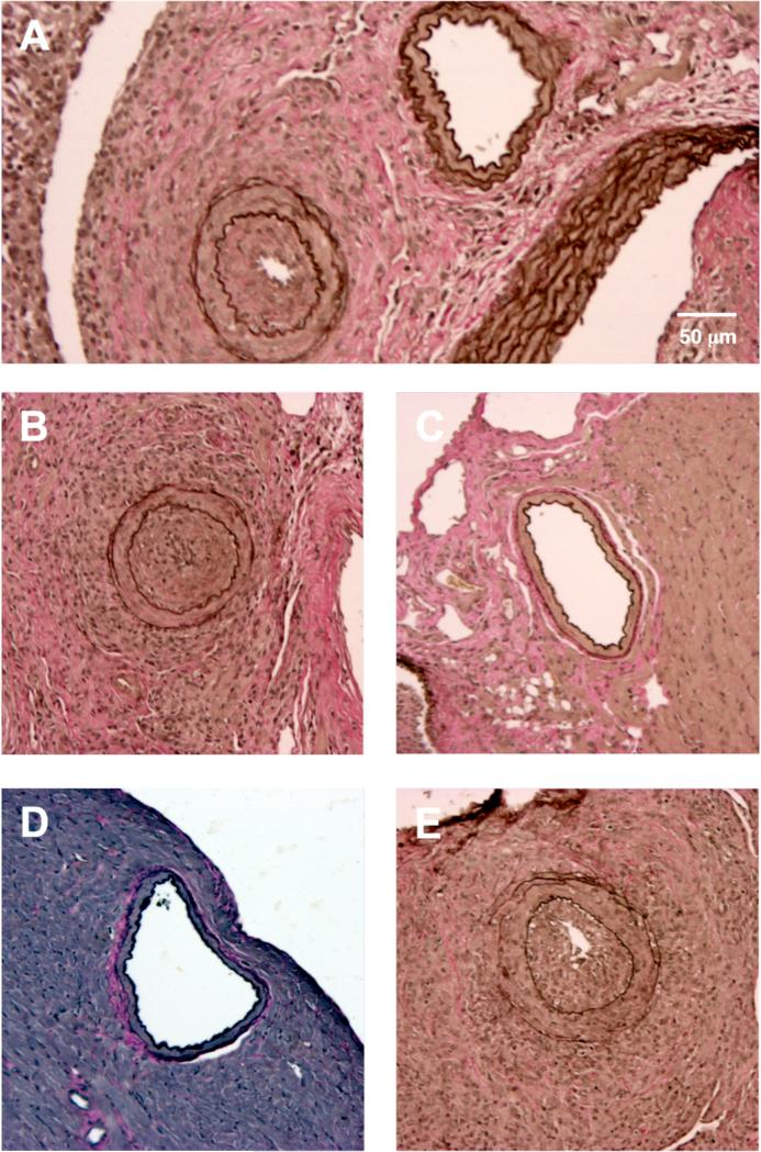
Figure 1. Representative cross-sections of coronary arteries of transplanted C3H cardiac allografts.
(A-B) Coronary arteries from B6.rag−/− recipients that received DSA (α-H2k IgG2a). Notably, severely occluded vessels were observed in close proximity to those that appeared completely normal and without neointimal thickening (A), which illustrates the heterogeneous nature of lesion formation. This is in comparison to (C), a widely patent coronary artery from a B6.rag−/− recipient that received no treatment. (D) A widely patent coronary artery from a B6.rag−/−γc−/− recipient that received no treatment in comparison to (E), a severely stenotic coronary artery from a B6.rag−/−γc−/− recipient that received DSA in addition to adoptively transferred (1.5 × 10⁶) B6 wild-type NK cells. A-E, elastin stain, scale bar in 1A equivalent in all other images (10x magnification). DSA, donor-specific antibody.

