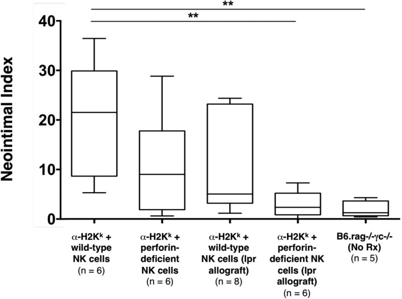
Figure 4. Effects of perturbations in cytolytic pathways on NK cell and DSA mediated chronic allograft vasculopathy.
In B6.rag−/−γc−/− recipients bearing C3H allografts that received DSA (α-H2k IgG2a) and perforin-deficient NK cells, CAV formation was minimally decreased, but not statistically significant in comparison to controls. Concurrently, in B6.rag−/−γc−/− recipients bearing C3H.lpr (Fas deficient) allografts that received DSA and wild-type NK cells, CAV formation again was minimally decreased, but not statistically significant in comparison to controls. Neither deficiency in perforin nor the Fas/FasL pathway alone altered CAV formation. However, a combined absence of these two pathways lead to a significant reduction in CAV (p = 0.0043) to a level that was the same as control animals that received no treatment (p = 0.662). Control groups indicated on this figure are the same as depicted in Figure 2 and are provided for comparative purposes. Annotations are as in Figure 2. CAV, chronic allograft vasculopathy; DSA, donor-specific antibody.

