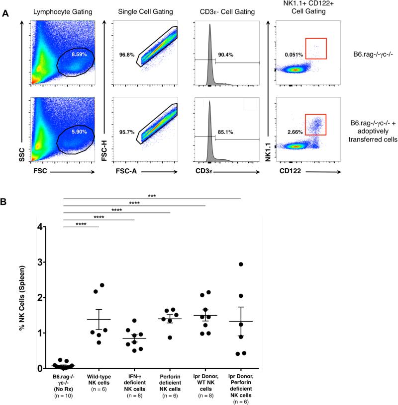
Figure 5. Adoptively transferred NK cells persist in B6.rag−/−γc− recipients at day 30 post-transplantation.
(A) Gating strategy to identify NK cells post-transplantation and adoptive transfer. A lymphocyte population was identified via forward / side scatter and further narrowed to single cells. Cells were then gated for those that were CD3ε-, CD122+ and NK 1.1+. Row 1 indicates representative flow plots from a B6.rag−/−γc−/− recipient that did not receive adoptively transferred cells. Row 2 indicates representative flow plots from a B6.rag−/−γc−/− that received adoptively transferred NK cells. (B) Whole spleens were obtained from recipients at the time of allograft recovery and analyzed with flow cytometry for the presence of NK cells. At baseline, B6.rag−/−γc−/− recipients have no mature NK cells. The percentage of NK cells in groups that received adoptively transferred cells was significantly higher than control animals (p < 0.001 for all groups). Error bars indicate the mean ± SEM. There was no significant difference in the percentage of cells between the treatment groups on statistical analysis using one-way ANOVA (p = 0.226).

