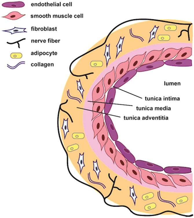
Fig. 1.

Anatomy of a blood vessel. Bloods vessels are comprised of 3 layers: the tunica intima, tunica media, and tunica adventitia. The innermost layer, the tunica intima, is comprised of a single layer of endothelial cells. The middle layer, the tunica media is predominately comprised of smooth muscle cells. The outermost layer, the tunica adventitia, consists of nerve fibers, fibroblasts, perivascular adipose tissue and collagen. Compared to smaller vessels (as depicted here), large vessels have increased tunica intima/media/adventitia thick- ness due to increased numbers of cells in each layer.
