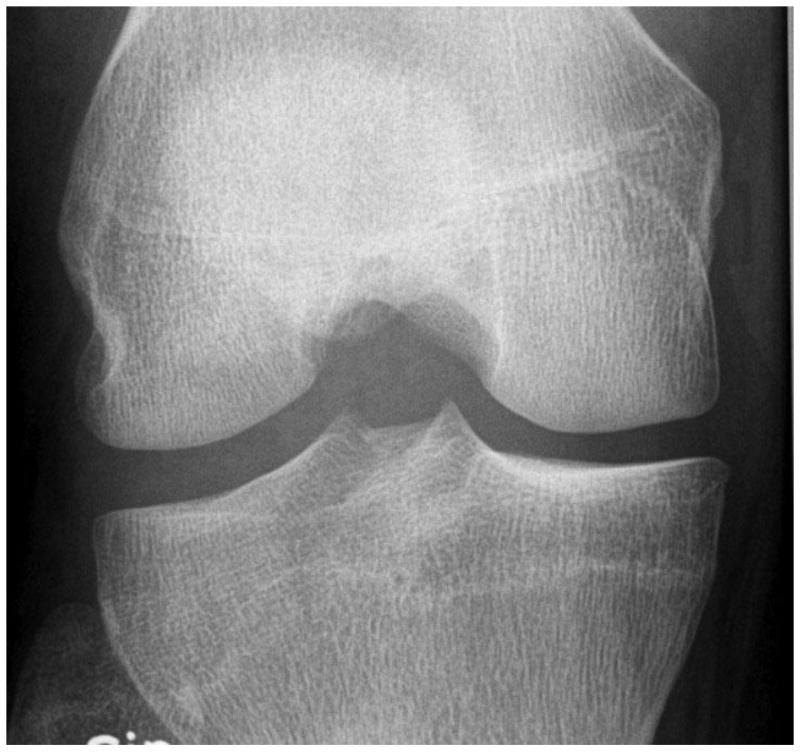
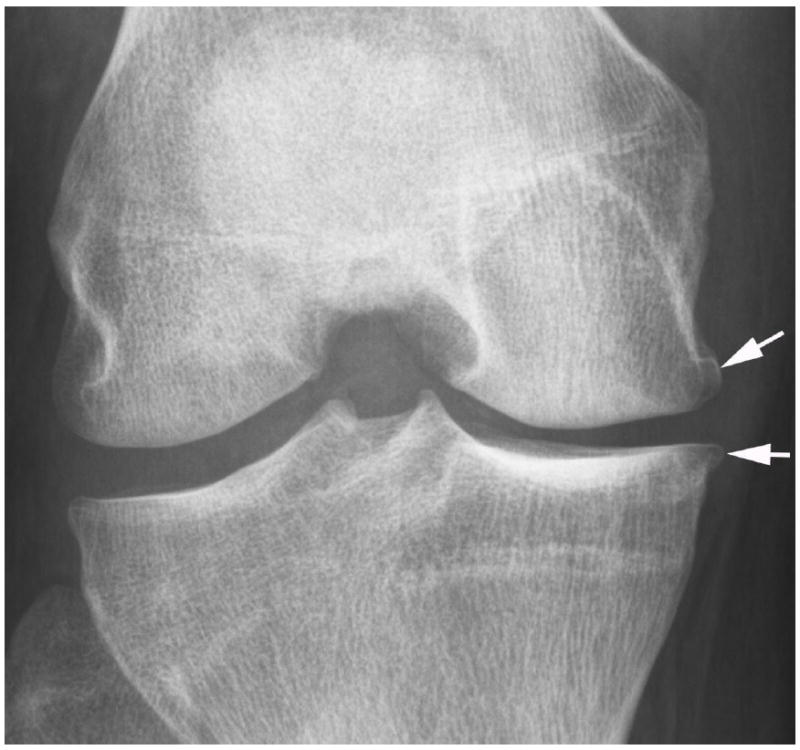
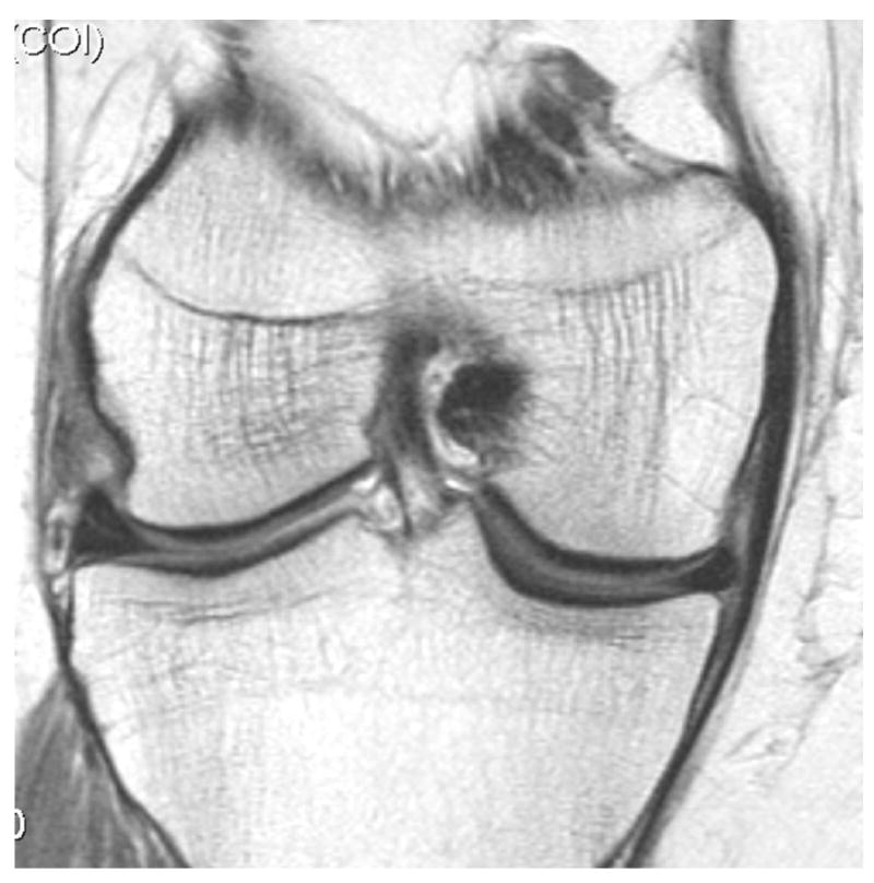
Fig. 2.


Development of radiographic osteoarthritis (OA) after partial meniscectomy. A. Baseline anterior–posterior radiograph of the knee shows physiologic joint anatomy without signs of OA. B. 12-month follow-up image shows definite radiographic OA with presence of medial tibial and femoral osteophytes at the joint margin consistent with radiographic OA Kellgren-Lawrence grade 2. C. Corresponding baseline MRI confirms absence of structural findings of OA. D. Follow-up image confirms partial meniscectomy with missing meniscal substance of the meniscal body (black arrow) and incident cartilage thinning (white arrows). An incident osteophyte is also depicted on the MRI (arrowhead).
