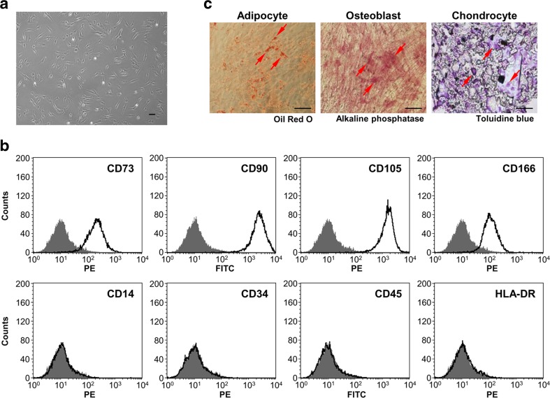
Fig. 1.
Characteristics of MSCs derived from human BM. a Representative morphological appearance of BM-MSCs. The cells exhibited a spindle-shaped or fibroblastic morphology. Scale bar at 100 μm. b Flow cytometric analysis of MSC surface markers. The expression of surface antigens was plotted against appropriate human IgG isotype controls (gray histograms). MSCs used in this study were positive for CD73, CD90, CD105, and CD166 and negative for CD14, CD34, CD45, and HLA-DR (clear histograms). c Differentiation of BM-MSCs. The cells were incubated for 14–21 days in the presence of specific differentiation agents for osteoblasts, chondrocytes, or adipocytes. Differentiation into the adipocyte lineage was demonstrated by staining with Oil-red O (red arrow), indicating intracellular lipid accumulation. Differentiation into osteoblast was demonstrated by staining with alkaline phosphatase (red arrow), indicating mineralization of the extracellular matrix. Differentiation into chondrocyte was demonstrated by staining with Toluidine Blue (red arrow), indicating the deposition of proteoglycans and lacunae formation. Scale bar at 100 μm

