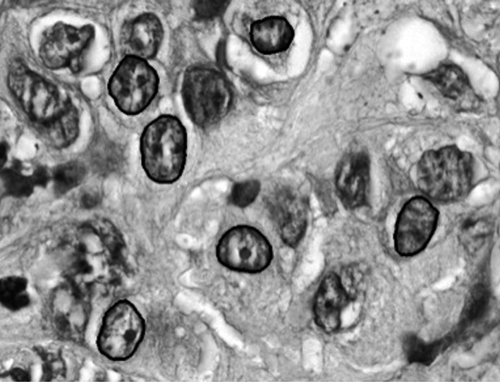
Figure 1.

Gray scale image of H&E slide of pancreatic carcinoma demonstrating segmentation of nuclei. A semiautomated imaging algorithm segments the nuclei from surrounding cytoplasm and artifacts. Within a segmented nucleus, each pixel is analyzed and mapped in a grid (x‐y plane) and analyzed with results seen in Table 2.
