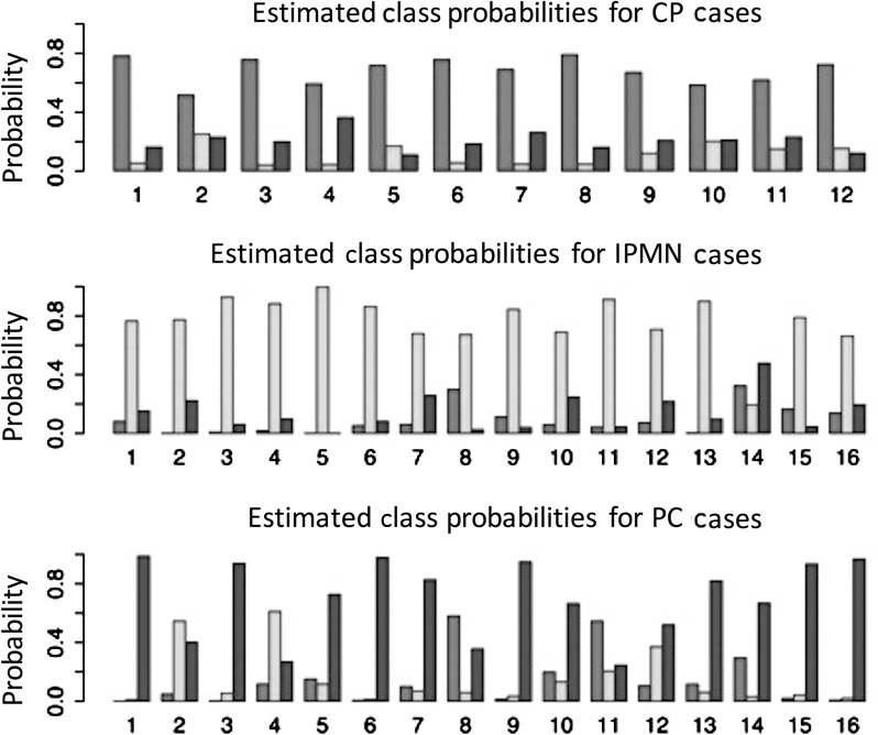
Figure 2.

The probability that a lesion was identified as CP (medium gray), IPMN (light gray), or PC (dark gray) is demonstrated for each lesion regardless of its true diagnosis. Proper lesion classification, as defined by the maximum probability of the three options, was achieved in 89.6% of lesions, with one IPMN and four PC misclassified. CP, chronic pancreatitis; IPMN, intraductal papillary mucinous neoplasms, PC, pancreatic carcinoma.
