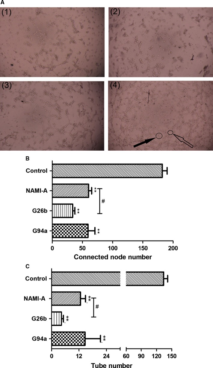
Figure 5.

The effect of ruthenium complexes on human umbilical vein endothelial cells (HUVEC) tube formation. HUVECs were seeded on Matrigel‐coated 96‐well plates and incubated in the diluted medium containing 20 μmol/L ruthenium complexes for 5 h at 37°C. (A) The images were captured under a phase contrast microscope at a magnification of 40× and observed the network‐like structures. (1) NAMI‐A; (2) G26b; (3) G94a; (4) control group. Solid black arrow stands for connected nodes, and feint arrow stands for tubes. (B) The connected node number of HUVEC; (C) The tube number of HUVEC. Values represent mean ± SD from three independent experiments. **P < 0.01 compared with the control group. G26b versus NAMI‐A group at the same dose, #stands for P < 0.05.
