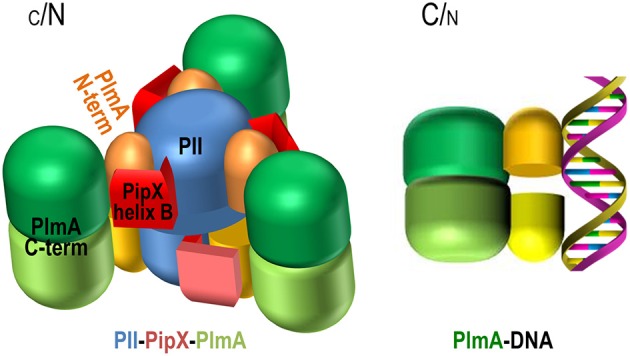
Figure 8.

Representation of the possible structure of the PII-PipX-PlmA complex and model for PlmA regulation according to C/N balance. PII trimers are blue, C-terminal helices of the PipX molecules (schematized in the extended conformation) are colored in two hues of red, and the N-terminal domains of PlmA dimers are colored yellow or orange, whereas the C-terminal domains are shown in two hues of green. The three-fold axis of the PII-PipX-PlmA complex is vertical and makes an angle of about 10° with the plane of the paper, exiting toward the viewer above the point of crossing with the paper. See text for more details.
