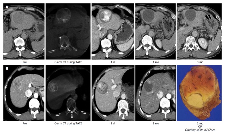
Figure 1.

Typical imaging finding after transarterial chemoembolization with drug-eluting bead. A: Nodular hepatocellular carcinoma (HCC) with arterial enhancement showed the total necrosis of HCC through follow-up computed tomography (CT) imaging; B: Nodular HCC showed nodular arterial enhancing viable portion within the partial necrosis of HCC in follow-up CT imaging. After operation, viable HCC in resected HCC showed the matched lesion in the CT imaging.
