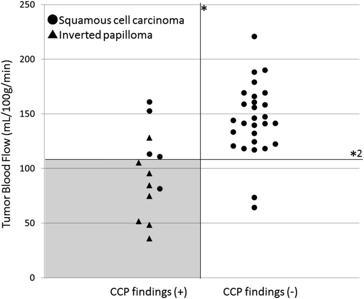
Figure 5.
Two-dimensional plot graph with the presence of convoluted ceribriform pattern (CCP) findings and tumour blood flow (TBF). Two-dimensional plot graph with the TBF values on the vertical axis and the presence of CCP findings on the horizontal axis was shown. By the combination use of CCP findings (*) and setting the range of 106–109 ml 100 g−1 min−1 at the threshold of TBF (*2), the highest diagnostic accuracy of 0.95 (39/41) with sensitivity of 0.97 (32/33) and specificity of 0.87 (7/8) was obtained. Most of the patients with IP can be successfully detected by using this threshold and the imaging findings of CCP with few overlaps with patients with squamous cell carcinoma (grey area).

