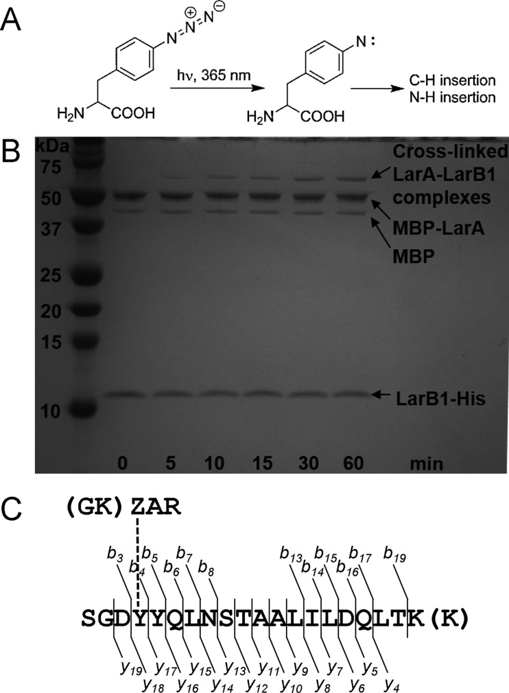
Figure 4.
Photocrosslinking of LarA to LarB1. (A) Structure of azidophenylalanine (AzF) and the nitrene that forms upon photolysis. (B) SDS–PAGE analysis showing that the band for LarA–LarB1 adducts grows as expected with the length of UV exposure. (C) Location of LarA cross-linked to LarB1 as determined by LC–MS/MS. The AzF incorporated into LarA is labeled Z. Amino acids in parentheses correspond to missed cleavages in the tryptic digestion. The observed b- and y-ions are shown, indicating that the cross-linking occurred to Tyr-28 of LarB1. Spectra corresponding to this figure are in Figure S9.

