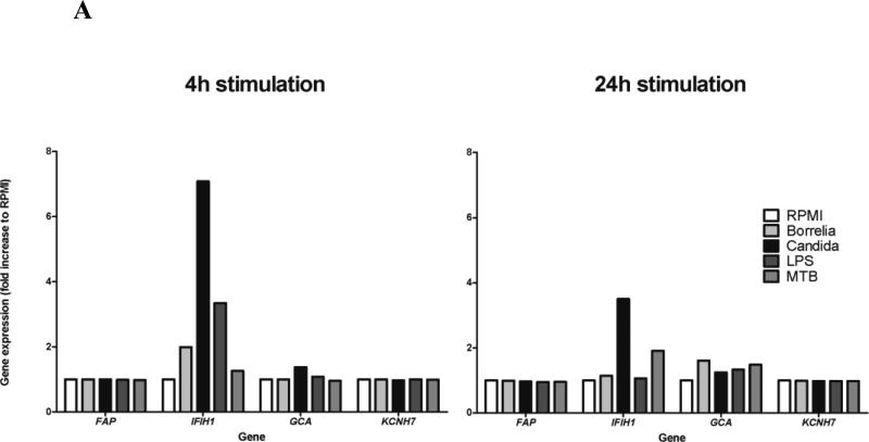
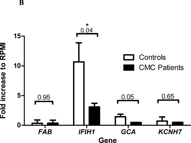
Figure 3. Transcriptional response of genes in the FAP-IFIH1-GCA-KCNH7 LD region to various microbial stimuli.
(A) Peripheral blood mononuclear cells (PBMCs) from healthy volunteers were stimulated for either 4 or 24 hours with Borrelia burgdorferi, Candida albicans, Escherichia coli-derived lipopolysaccharide (LPS), or Mycobacterium tuberculosis (MTB). Gene expression was measured using microarrays and normalized to the control RPMI condition (untreated). (B) Gene expression (Mean ± SD) in PBMCs of healthy controls (n=3) and patients suffering from chronic mucocutaneous candidiasis (CMC) (n=2) were stimulated with C. albicans for 4 hours. P values were calculated using the Welch-corrected t test.


