Figure 3.
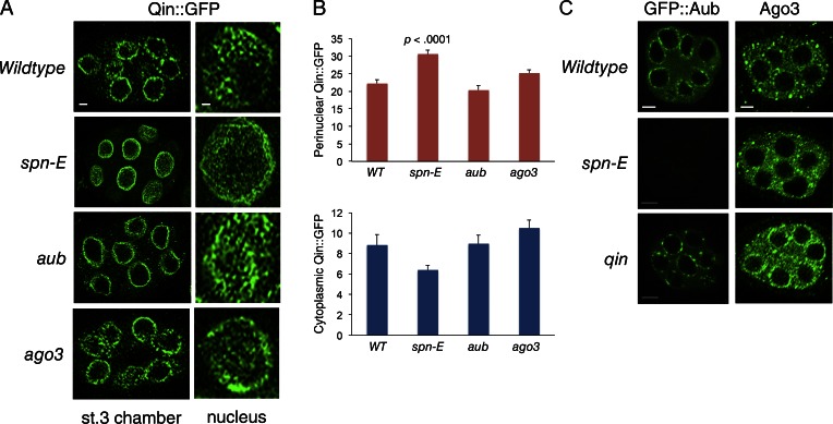
Spn-E regulates localization of Qin and PIWI proteins. (A) Qin::GFP localization in stage 3 egg chambers of wild type and mutants, as indicated. (Left) Section through one egg chamber. Bar, 5 µm. (Right) Tangent planar views of a single nurse cell nucleus from each genotype. This plane images the perinuclear localization of Qin::GFP. Bar, 1 µm. Shown are representative egg chambers from a total number imaged and analyzed of n = 50 (wild type), n = 22 (spn-E), n = 16 (aub), and n = 12 (ago3). (B) Quantification of Qin::GFP fluorescence intensity in defined regions of nurse cells. Shown are mean intensities detected for perinuclear Qin::GFP and peripheral cytoplasmic Qin::GFP. Error bars are SEM; two-tailed t tests were done to compare the difference in intensities between wild type (WT) and each mutant. The result of the only t test returning significance is shown. Replicate number for each analysis (bar) ranged from 12 to 18. (C) GFP::Aub localization in stage 3 egg chambers of wild type and mutants, as indicated. Ago3 protein localization as detected by anti-Ago3 in stage 3 egg chambers of wild type and mutants, as indicated. Bars, 5 µm.

