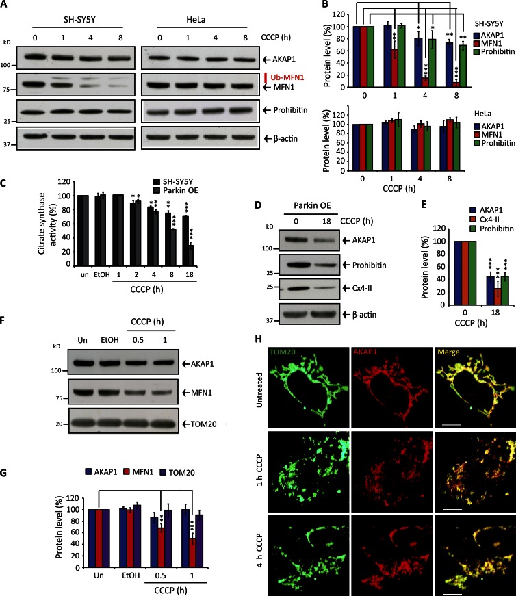
Figure 2.
Mitochondrial depolarization does not trigger AKAP1 degradation or release from the OMM. (A and B) AKAP1, MFN1, and Prohibitin protein levels in 0–8 h depolarized SH-SY5Y or HeLa cells. Quantification was corrected for β-actin. (C) Citrate synthase activity adjusted for protein (BCA) after 0–18-h CCCP treatment in SH-SY5Y cells expressing endogenous and overexpressed Parkin. (D and E) AKAP1, Prohibitin, and Cx4-II protein levels in Parkin-overexpressing SH-SY5Y cells incubated with CCCP for 18 h. Quantification was corrected for β-actin. (F and G) AKAP1, MFN1, and TOM20 protein levels and quantification in mitochondria isolated from CCCP-treated SH-SY5Y cells. un, untreated. (H) Immunocytochemistry demonstrating AKAP1 colocalization with TOM20 in SH-SY5Y cells during 0–4-h CCCP treatment. Pearson coefficients as follows: untreated = 0.84 ± 0.03; 1-h CCCP = 0.85 ± 0.02; 4-h CCCP = 0.83 ± 0.01. Bars, 20 µM. Each experiment was repeated at least three separate times, and error bars indicate SDM. *, P < 0.05; **, P < 0.01; ***, P < 0.001.

