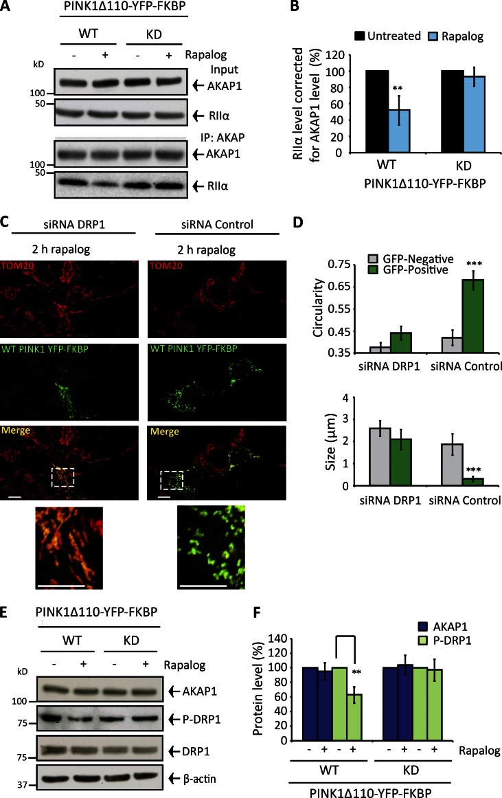
Figure 4.
Controlled targeting of PINK1 to the OMM disrupts the AKAP1–PKA axis and drives fission via DRP1. (A and B) SH-SY5Y cells transfected with WT or KD PINK1Δ110-YFP-FKBP/FRB-Fis1 (48 h) and treated with rapalog for 1 h. (A) AKAP1-RIIα CoIP. (B) RIIα abundance corrected for AKAP1 and normalized to 100% in untreated samples. (C and D) SH-SY5Y cells 72 h drp1- or control-silenced and transfected with WT or KD PINK1Δ110-YFP-FKBP/FRB-Fis1 for 48 h. Mitochondrial morphology (TOM20-stained) and quantification after 2-h rapalog treatment. (E and F) AKAP1, DRP1, and phospho–serine 637 DRP1 levels in SH-SY5Y cells expressing WT or KD PINK1Δ110-YFP-FKBP/FRB-Fis1 (48 h) with or without 2-h rapalog treatment. Phospho-DRP1 adjusted for DRP1/β-actin and AKAP1 for β-actin. Experiments were repeated at least three times, and error bars indicate SDM. Bars: 20 µM; (insets) 10 µM. **, P < 0.01; ***, P < 0.001.

