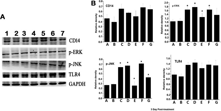
Fig. 10.

Western blotting analyzed the expression of TLR4-related protein in the lung tissue after MSC transplantation for 3 days. a Expression of CD14, TLR4, ERK, and JNK were evaluated by immunoblotting using specific antibodies. GAPDH is used as the control. 1: controls, 2: MSCs, 3: H9N2, 4: H9N2 + McCoy, 5: H9N2 + MSCs, 6: H9N2 + Physiological saline (1 day following) 7: H9N2 +MSCs (1 day following). b Responses were quantified by densitometry and normalized to the expression of GAPDH. Densitometry data are shown as mean ± SD. *Response that is significantly different from the control (p < 0.05). ▲Response that is significantly different from the H9N2 infected mice (p < 0.05)
