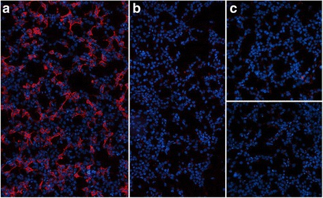
Fig. 8.

MOG-IgG1 as detected in the fixed-cell CBA. a, b Binding of serum IgG1 antibodies (from a patient with recurrent optic neuritis) to HEK293 cells transfected with human full-length MOG (a), but not to mock-transfected HEK293 cells (b). c Negative control serum (from a patient with RRMS) binding neither to the MOG-transfected cells (upper panel) nor to the mock-transfected control cells (lower panel). Bound patient IgG1 was detected by successive incubation with an unlabeled sheep anti-human IgG1 secondary antibody and an AlexaFluore®568-labeled donkey anti-sheep IgG antibody (red fluorescence). Cell nuclei were stained with 4’,6-diamidino-2-phenylindole (blue fluorescence)
