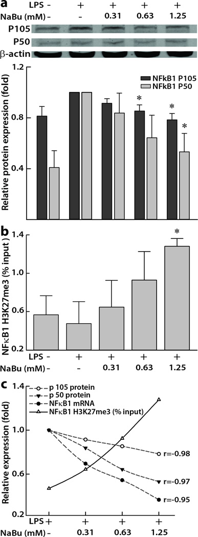
Figure 2. Concentration-dependent effects of NaBu on NFκB1 in human colon epithelial cells.
(a) Representative immunoblots showing the suppression of total cellular NFκB1 subunits p105 and p50. Densitometric analyses showing relative protein expressions normalized to β-actin proteins and expressed as mean ± SEM (n = 3). *p < 0.05, compared with LPS (10 ng/mL) treated positive control. (b) Histone 3 lysine 27 tri-methylation (H3K27me3) changes at promoter regions of NFκB1 in cells treated with different concentrations (0.31 to 1.25 mM) of NaBu for 5 h. Data points represent the input ± SEM (n ≥ 4). *p < 0.05, compared with LPS control.

