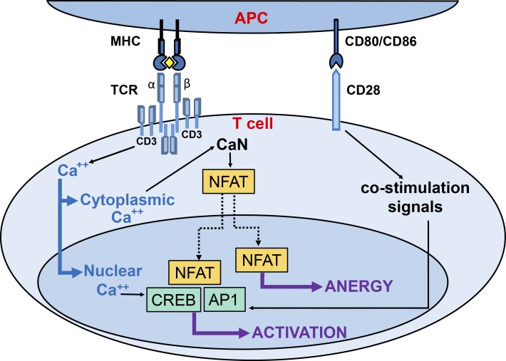
Figure 7.
Differential role of spatially distinct calcium signals in the human T cell immune response. Schematic illustration of the roles of nuclear and cytoplasmic calcium signals in the decision of stimulated T cells to mount a proliferative immune response or develop tolerance. In the immunological synapse, the stimulation of TCR and CD28 by the corresponding receptors on the antigen-presenting cell (APC) trigger the activation of different signaling pathways that lead to the induction of the immune response. After being activated by binding to the major histocompatibility complex (MHC)/antigen complex (the yellow rhombus represents the antigen), CD3 subunit of TCR induces an intracellular calcium increase, which activates CaN and NFAT in the cytoplasm and CREB and AP1 in the nucleus. The selective blockade of nuclear calcium signaling inhibits CREB activation but not NFAT translocation to the cell nucleus. As a consequence, T cell activation is blocked, and the immune response is redirected toward an anergy-like state.

