Figure 6.

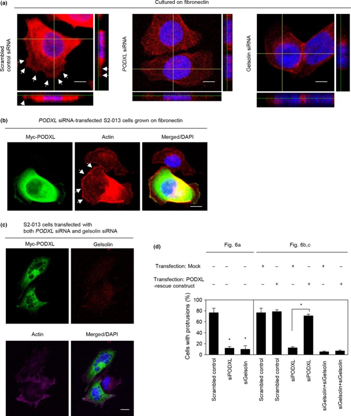
Roles of podocalyxin‐like protein (PODXL) and gelsolin in forming cell protrusions. (a) Confocal Z stack shows phalloidin‐labeled peripheral actin structures (red) and DAPI‐labeled nuclei (blue) in scrambled control siRNA‐transfected S2‐013 pancreatic ductal adenocarcinoma cells, PODXL siRNA‐transfected S2‐013 cells, or gelsolin siRNA‐transfected S2‐013 cells grown on fibronectin. Arrows, peripheral actin structures in cell protrusions. The lower and right panels in the confocal Z stack show a vertical cross‐section (yellow lines) through the cells. Bar = 10 μm. (b) Confocal immunofluorescence microscopic images. A myc‐tagged PODXL‐rescue construct was transfected into S2‐013 cells that had been transfected with PODXL siRNA. After 48 h, cells were incubated on fibronectin. Cells were stained with anti‐myc antibody (green) and phalloidin (red). Arrows, cell protrusions reproduced by exogenous PODXL in PODXL siRNA‐transfected cells. Blue, DAPI staining. Bar = 10 μm. (c) Confocal immunofluorescence microscopic images. A myc‐tagged PODXL‐rescue construct was transfected into S2‐013 cells that had been transfected with PODXL siRNA and gelsolin siRNA. After 48 h, the cells were incubated on fibronectin. The cells were stained with anti‐myc antibody (green), anti‐gelsolin antibody (red), and phalloidin (violet). Blue, DAPI staining. Bar = 10 μm. (d) Quantification of data shown in (a–c); the values represent the number of cells with fibronectin‐mediated cell protrusions in which peripheral actin structures were increased. All cells in four fields per group were scored. Data are derived from three independent experiments. Columns, mean; bars, SD. *P < 0.001 compared with corresponding PODXL siRNA‐transfected S2‐013 cells that were transfected with mock vector (Student's t‐test).
