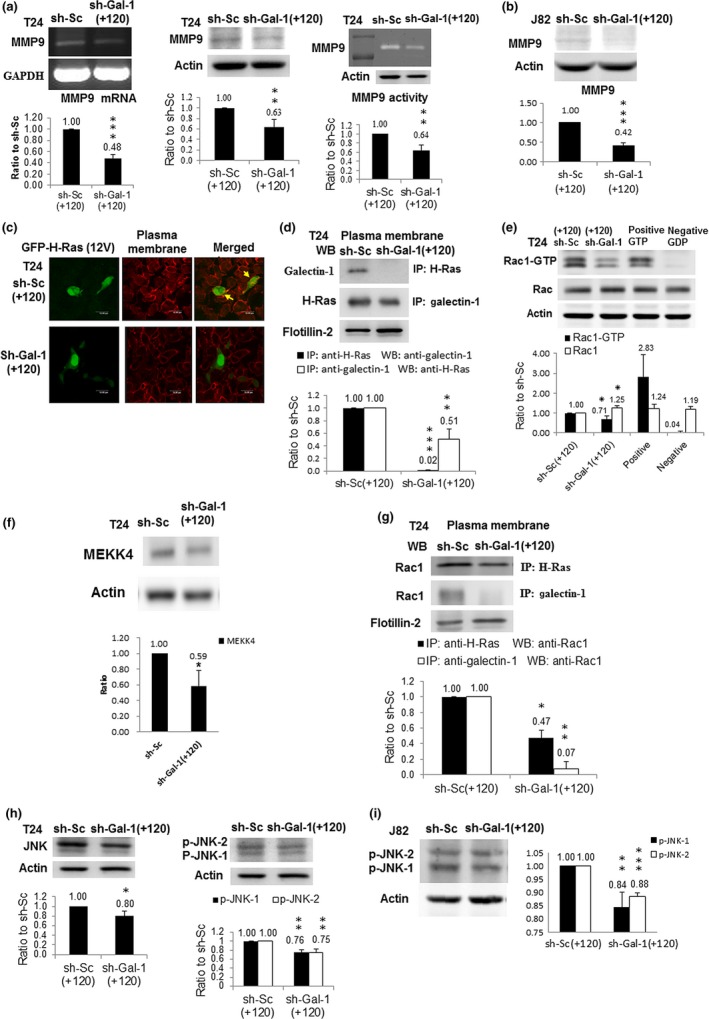
Figure 2.

Impacts of galectin‐1 (Gal‐1) silencing on the Gal‐1‐mediated signaling pathway. (a) MMP9 protein (left panel) and mRNA (middle panel) amounts in T24 cell clones with knockdown at +120 nt (shGal‐1(+120)) measured by Western immunoblotting and RT‐PCR, respectively. MMP9 activities (right panel) in shGal‐1(+120) T24 cells were evaluated by zymography. The blot in each figure was the typical result of at least three independent studies. The diagram (ratio [mean ± SD]) under each blot shows the differential protein expression that was expressed as the ratio of normalized intensity (observed protein/actin) of sh‐Gal‐1(+120) cells divided by that of scrambled sh‐Sc(+120) cells. The number above each bar is the average ratio. (b) MMP9 protein level in shGal‐1(+120) J82 cells. (c) Confocal microscopic image of shGal‐1(+120) T24 cells, typical of three independent experiments. shGal‐1(+120) and sh‐Sc(+120) T24 cells were transfected with H‐Ras expression vector (pMSCV‐H‐Ras(12V)‐GFP). H‐Ras protein is shown in green and the cell membrane is stained in red in T24 sh‐Sc(+120) cells. Yellow (arrow), shown on the cell membrane in the merged image, indicates that H‐Ras was located on cell membrane of T24 sh‐Sc(+120) cells. However, the cell membrane remained red in the merged image of T24 sh‐Gal‐1(+120) cells, indicating that H‐Ras did not appear on the cell membrane because of Gal‐1 knockdown. (d) Interaction of Gal‐1 with H‐Ras in plasma membranes of shGal‐1(+120) cells. Protein lysate from purified plasma membrane immunoprecipitated (IP) with antibodies against H‐Ras and Gal‐1 proteins present in the immunocomplex was detected by western immunoblotting and vice versa. (e) Rac1 protein level in shGal‐1(+120) T24 cells. Protein lysates were collected for Western immunoblotting (WB). GTP or GDP‐incorporated protein lysates were used as positive or negative controls. (f) MEKK4 protein amount in shGal‐1(+120) T24 cells. (g) Interaction of Rac1 with H‐Ras or Gal‐1 in plasma membranes of shGal‐1(+120) cells. Protein lysate from purified plasma membrane was immunoprecipitated with antibody against H‐Ras or Gal‐1 protein and Rac1 protein present in the immunocomplex was detected by Western immunoblotting. Flotillin‐2 was the loading control. (h) JNK (left panel) and phosphorylated (p‐)JNK) (right panel) protein levels in shGal‐1(+120) T24 cells. (i) p‐JNK protein amount in shGal‐1(+120) J82 cells. *P ≤ 0.05, **P ≤ 0.01, ***P ≤ 0.001.
