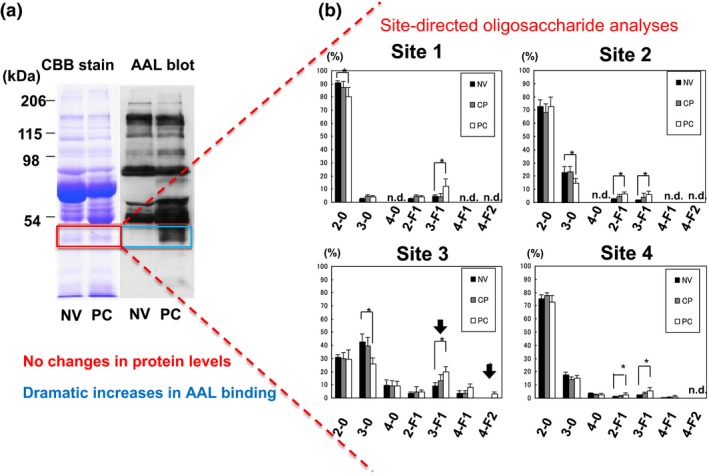
Figure 1.

Identification of fucosylated haptoglobin and site‐directed oligosaccharide analyses of haptoglobin. (a) An approximately 40‐kDa protein was specifically fucosylated in the sera of pancreatic cancer (PC) patients, whereas its protein level was almost the same. This figure is adapted from Okuyama et al.17 with slight modification, with permission from Wiley. (b) Site‐specific oligosaccharide analyses with mass spectrometry on purified oligosaccharides from sera of normal volunteers (NV), patients with chronic pancreatitis (CP), and those with PC. Site 3 is susceptible to fucosylation, and the glycan structure 4F‐2 was specifically detected at this site. These data adapted from Nakano et al.18 with slight modification, with permission from Wiley. Numbers on the x‐axis in each figure part indicate branching on N‐glycans; F indicates the number of fucose molecules attached on each N‐glycan. n.d., not detected.
