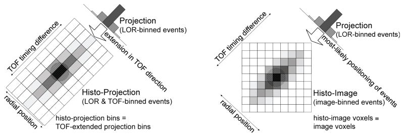
Figure 1.
Comparison of TOF data partitioning (for 2D case) for one angle binned into histo-projections (left) or into histo-images with the DIRECT approach (right). Histo-projections can be described as an extension of non-TOF projections (radial bins) along the TOF direction (time bins); the sampling intervals relate to the projection geometry and TOF resolution, while histo-image sampling is given by the voxel geometry of the reconstructed image. (This figure was reproduced from (Nuyts and Matej, 2015) (Figure 13.13) with permission by the IAEA.)

