
Figure 2.
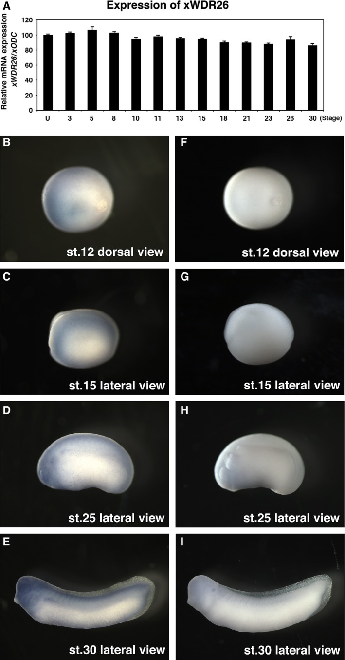
Expression of xWDR26 during Xenopus embryogenesis. (A) Quantitative RT‐PCR revealed temporal expression of xWDR26. Numbers under lanes indicate developmental stages; U, unfertilized eggs. The value obtained for xWDR26 was normalized to the level of xODC (ornithine decarboxylase). The value of unfertilized eggs was set to 100 and other values were computed. Error bars represent standard deviation of the mean in three experiments. Statistical significances of xWDR26/xODC between unfertilized eggs and other stages were determined by Mann–Whitney U test. P > 0.05 (until stage 15, and stage 26), P < 0.01 (after stage 18, except for stage 26). (B–I) Whole‐mount in situ hybridization. (B–E) Anti‐sense RNA probe of xWDR26 cDNA fragment was used. (F–I) Sense RNA probe of xWDR26 cDNA fragment was used. (B and C). Expression of xWDR26 gradually localizes to anterior neural region (B, stage 12; C, stage 15). (D and F) xWDR26 is strongly expressed in the anterior neural region (D, stage 25; E, stage 30).