
Figure 3.
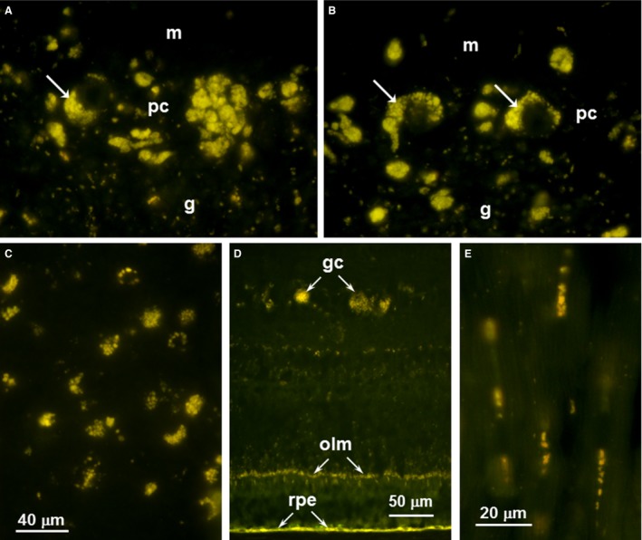
Fluorescence micrographs of cryostat sections the cerebellum (A and B), cerebral cortex (C), retina (D) from Dog A and heart ventricular wall (E) from Dog C. In the cerebellum, storage body accumulation was most abundant in the Purkinje cell layer (pc) with some but not all of the material within cells that could be identified as Purkinje cells (arrows in A and B). There were lesser accumulations of this material in the molecular (m) and granular (g) layers. In the cerebral cortex (B), the storage material was widely distributed throughout most of the tissue. In the retina (C), the most prominent accumulations of autofluorescent material were in the ganglion cells (gc) and along the outer limiting membrane (olm). The retinal pigment epithelium (rpe) also contained substantial amounts of material with similar fluorescence properties, but since the accumulation of such material occurs during normal aging, the presence of this material in the rpe is not diagnostic for NCL. Clusters of autofluorescent inclusions were present in the heart muscle fibers sections in longitudinal orientation (E).