
Figure 2.
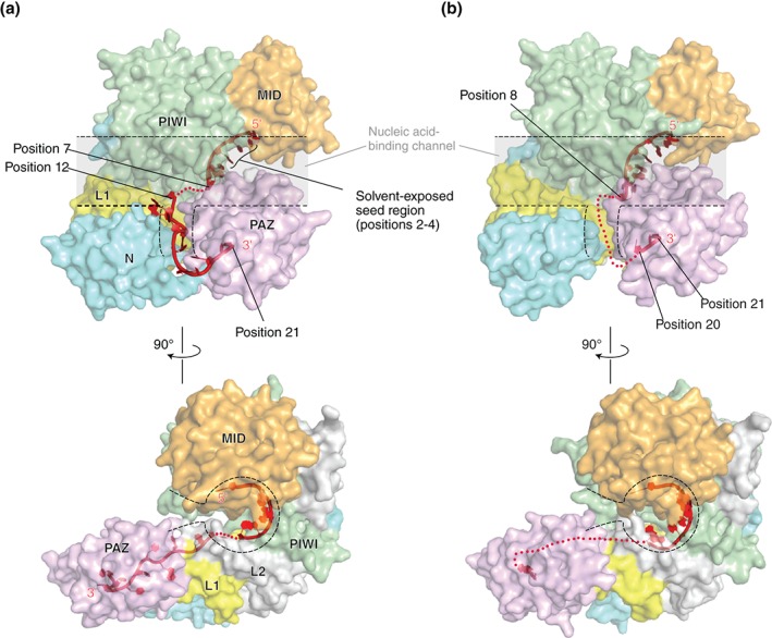
Structures of the RISC of human Argonaute2 (a: PDB ID: 4W5N) and Argonaute1 (b: PDB ID: 4KXT). The transparent surface models of hAGOs are drawn with the same color codes as in Figure 1. The linkers, L1 and L2, are colored in yellow and gray, respectively. The nucleic acid‐binding channels are highlighted with dotted lines. The guide RNA (red) is depicted as a ribbon model. The disordered parts of the guide are shown as dotted lines.