
Figure 7.
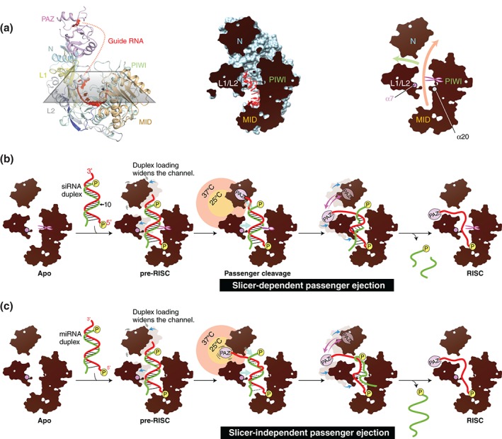
Two model mechanisms of passenger ejection. (a) Y‐shaped nucleic acid‐binding channel of AGO protein. Left: The crystal structure of hAGO2‐RISC (PDB ID: 4OLA) is cut with a section. The color codes of hAGO2 are the same as in Figure 1. Middle: The hAGO2 (light blue) and the bound guide RNA (red) are shown as a surface and a ball‐and‐stick model, respectively. The section area is colored in black. Right: The branched channels. The main channel and the branch are shown by orange and green arrows, respectively. The catalytic site is indicated by scissors. The α7 and α20 are drawn as spheres. (b) Model of the slicer‐dependent passenger ejection. The siRNA duplex composed of a guide (red) and a passenger (green) is loaded onto hAGO2 to form the pre‐RISC. The 5′ monophosphate is shown as a yellow sphere. The N and L1/L2 domains move outward (blue arrow) to expand the width of the nucleic acid‐binding channel. Only the guide RNA is anchored at its 5′ monophosphate and the sugar‐phosphate backbone in the seed region. The loaded siRNA duplex is pushed by the N and L1/L2 domains (cyan arrows) and squeezed out upon the passenger cleavage. The thermal dynamics of the PAZ domain shakes the 3′ end of the guide strand (pink arrows), which facilitates the ejection of the cleaved passenger strand. The territories of the PAZ domain at 25 and 37°C are drawn in a yellow and salmon circles, respectively. The guide 3′ end of siRNA duplex is positioned outside a territory in which the PAZ domain can reach at 25°C. Destabilized base pairs are depicted as dotted line. (c) Model of the slicer‐independent passenger ejection. The guide 3′ end is positioned within a territory that the PAZ domain can reach at 25°C. Because of its thermodynamic instability, the miRNA duplex is heavily distorted by inward pressure.