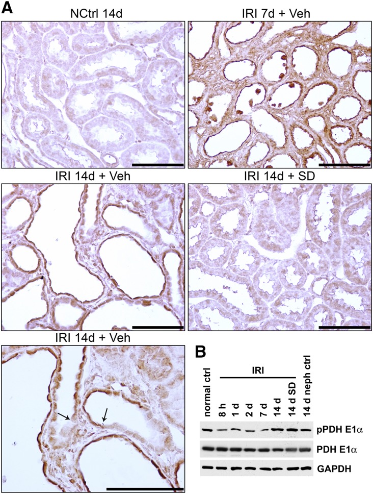
Figure 8.
Phosphorylation of pyruvate dehydrogenase in kidney tissue and tubules after IRI. (Upper panel) IHC for phosphopyruvate dehydrogenase E1α (pPDHE1α) in nephrectomy control, kidneys 7 and 14 days after IRI, and kidney with SD208 treatment 14 days after IRI. Arrows point to sharp transitions between pPDHE1α staining in atrophic epithelium and adjacent thicker differentiated epithelium with weak or no staining. Scale bar, 100 μm. (Lower panel) Western blotting of kidney lysates for pPDHE1α and PDHE1α in the time course and protocol outlined for Figure 2A. ctrl, Control; GAPDH, glyceraldehyde-3-phosphate dehydrogenase; NCtrl, nephrectomized control; neph, nephrectomy; Veh, vehicle.

