Figure 9.
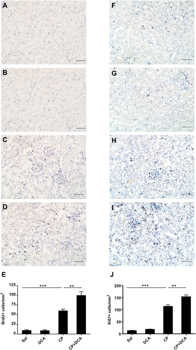
DCA enhances cellular proliferation in the kidneys during CP-induced AKI. Representative photomicrographs (magnification ×100) of BrdU staining in the kidneys of (A) Sal, (B) DCA, (C) CP, and (D) CP and DCA cotreated mice treated as outlined in the CP-induced AKI model section in the Concise Methods. BrdU-positive cells are stained brown. Bar=50 µm. (E) Quantitative analysis of BrdU-positive cells. Representative photomicrographs (magnification ×100) of Ki67 staining in the kidneys of (F) Sal, (G) DCA, (H) CP, and (I) CP and DCA cotreated mice treated, as outlined in the CP-induced AKI model section in the Concise Methods. (J) Quantitative analysis of Ki67-positive cells. Results are expressed as mean±SEM, n=8 animals per group ***P<0.001; **P<0.01.

