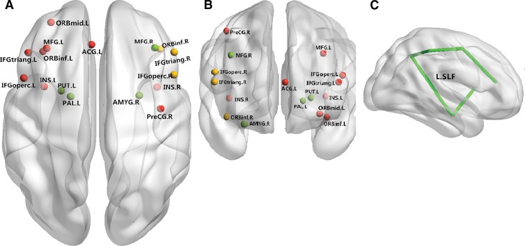
Figure 3:
Neural networks involved in BD mainly include the inferior frontal cortex and limbic areas. Both anatomic and functional changes are shown in the, A, superior view and, B, anterior view. Red spots and yellow spots represent changes in gray matter volume and function, respectively; green spots indicate regions with both functional and anatomic changes. C, Main white matter bundles (green line), with changes of integrities revealed by DT imaging. ACG.L = left anterior cingulate gyrus; AMYG.R = right amygdala; IFGoperc.L = left inferior frontal gyrus, pars opercularis; IFGoperc.R = right inferior frontal gyrus, pars opercularis; IFGtriang.L = left inferior frontal gyrus, pars triangularis; IFGtriang.R = right inferior frontal gyrus, pars triangularis; INS.L = left insula; INS.R = right insula; L.SLF = left superior longitudinal fasciculus; MFG.L = left middle frontal gyrus; MFG.R = right middle frontal gyrus; ORBinf.L = orbital part of left inferior frontal gyrus; ORBinf.R = orbital part of right inferior frontal gyrus; ORBmid.L = orbital part of left middle frontal gyrus; PAL.L = left lenticular nucleus, pallidum; PreCG.R = right precentral gyrus; PUT.L = left lenticular nucleus, putamen.

