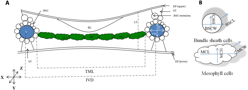
Fig 1. A schematic representation of a rice leaf shows different cell types and three different leaf dissection axes; used to calculate cell size, number, and volume.
For ease of viewing, bulliform and epidermal cells are not shown in detail. (A) Rice leaf transverse section. The position of major leaf cells: mesophyll cell (MC, coloured green), bundle sheath cells (BSC, coloured white), vein (V, coloured blue), bulliform cells (BL), stone cells (ST), and epidermal layers. In rice, inter-veinal distance is filled by a number of elliptic mesophyll cells. Veins are surrounded by a wreath of bundle sheath cells and crowned by bundle sheath cell extension above the main circle. Large bulliform cells are present only at the adaxial side of the leaf. Stone cells are present at both the abaxial and adaxial end of the vein. X, Y and Z represent the three growth axes where, X represents the leaf lateral axis, Y represents the leaf abaxial-adaxial axis, and Z represents the leaf longitudinal or the proximo-distal axis. The long axis of the mesophyll cell is perpendicular to the vein axis. The long axis of bundle sheath cell is parallel to the vein axis and perpendicular to the mesophyll cell. LT = Leaf thickness; IVD = Inter-veinal distance; TML = Total mesophyll length in inter-veinal space; VW = Vein width; VH = Vein height. (B) Mesophyll and bundle sheath cell parameters, measured along X, Y, and Z. MCL = mesophyll cell length; MCH = mesophyll cell height; MCW = Mesophyll cell width; and BSCW = Bundle sheath cell width; BSCH = Bundle sheath cell height; and BSCL = Bundle sheath cell length.

