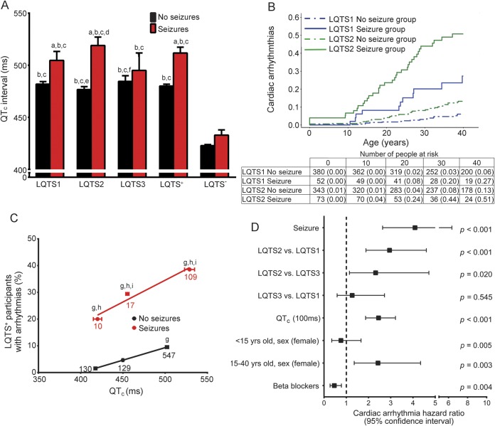
Figure 4. LQTS+ participants with seizures exhibited more severe ECG manifestations.
(A) Seizure cohort exhibited longer QTc durations (analysis of variance with Tukey test: LQTS− and LQTS+ with and without seizures, p < 0.001; LQTS1, 2, and 3, and LQTS− with and without seizures, p < 0.001; ap < 0.05 vs genotype no seizure; bp < 0.05 vs LQTS− no seizure; cp < 0.05 vs LQTS− seizure; dp < 0.05 vs LQTS1 no seizure; ep < 0.05 vs LQTS1 seizure; fp < 0.05 vs LQTS2 seizure). (B) Higher cumulative probability of cardiac arrhythmias in LQTS1 and LQTS2 participants with vs without seizures (log rank p < 0.001). (C) Within the same QTc window, there was a higher percentage of LQTS+ seizure participants who developed arrhythmias (n = participants; Cochran-Mantel-Haenszel test: LQTS+ no seizure; p < 0.001; LQTS+ seizure, p = not significant. Chi square: p < 0.001; gp < 0.05 vs LQTS+ no seizure; normal QTc; hp < 0.05 vs LQTS+ no seizure; borderline QT prolongation; ip < 0.05 vs LQTS+ no seizure; QTc prolongation. LQTS− no seizure; R2 = 0.9999, slope = 0.0939. LQTS+ seizure, R2 = 0.9568, slope = 0.1631). (D) Multivariate analysis indicated seizures were the strongest independent positive predictor of arrhythmias. LQTS = long QT syndrome; QTc = corrected QT interval.

