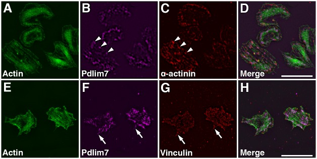
Fig 5. Organization of Pdlim7, actin, and actin-associated proteins during platelet shape change.
SIM images of thrombin activated WT platelets displaying intermediate spreading stages. Triple immunofluorescence staining determines the distribution of Pdlim7 in context of the cytoskeletal protein α-actinin and the focal adhesion protein vinculin. Top row shows SIM images of platelets immunostained for (A) F-actin (green), (B) Pdlim7 (magenta), (C) α-actinin (red), and (D) Merge. Bottom row shows SIM images of WT platelets immunostained for (E) F-actin, (F) Pdlim7 (magenta), (G) vinculin (red), and (H) Merge. Arrowheads in panel B and C indicate co-localization of Pdlim7 with α-actinin along actin stress fibers, and arrows in panel F and G point to Pdlim7 distribution in filopodia, just proximal of and abutting vinculin-rich adhesion sites. Scale bar = 5μm.

