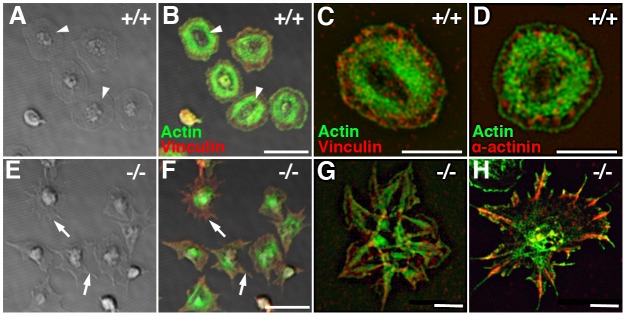
Fig 7. Loss of Pdlim7 disrupts normal actin organization in activated platelets.
DIC and confocal images (left hand panel, A, B, E, F) taken of WT and Pdlim7-/- platelets stimulated with thrombin, spread on glass, and stained for F-actin (green) and vinculin (red). 45 minutes after stimulation, WT platelets were fully spread and displayed an organized F-actin fiber ring (arrowhead, A, B). In contrast, Pdlim7-/- platelets did not spread, revealed unorganized actin accumulation in the center, and exhibited filopodia-like protrusions with low actin content (arrow, E, F). After completion of spreading, in WT platelets vinculin accumulates along a circle at the microfilament tips, whereas in Pdlim7-/- platelets it appears to be distributed throughout the platelet body and filling the filopodia-like protrusions (B, F). SIM images (right hand panel, C, D, G, H) show increased detail of actin (green) and vinculin (red) (C, G) as well as actin (green) and α-actinin (red) (D, H) distribution in fully spread WT and Pdlim7-/- platelets. Scale bar (A, B, E, F) = 5μm. Scale bar (C, D, G, H) = 2.5μm.

