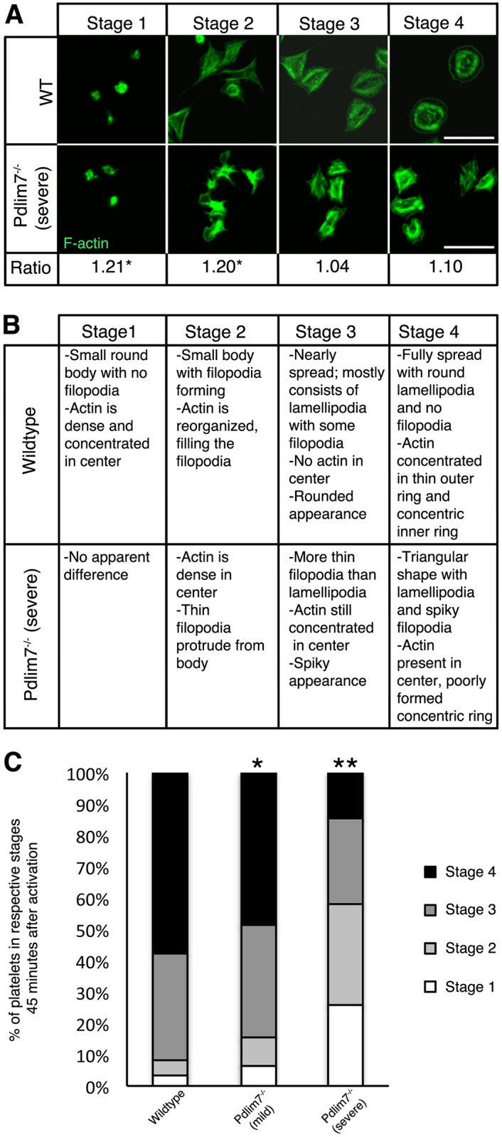
Fig 8. Spreading and F-actin dynamics are altered in activated Pdlim7-/- platelets.

Platelets from WT and Pdlim7-/- mice were thrombin stimulated and spread on glass for 45 minutes. Fixed platelets were immunostained for F-actin (green), imaged by confocal microscopy, and staged according to the following scheme: stage 1 (resting); stage 2 (filopodia only); stage 3 (filopodia and lamellipodia); and stage 4 (fully spread). (A) Representative images of Pdlim7-/- platelets presenting a severe phenotype reveal that F-actin remains disorganized and concentrated in the center of the platelets as compared to WT controls. Quantification of the epi-fluorescence intensities of the phalloidin signal of 100 individual platelets in each of the 4 stages and calculation of the ratios of Pdlim7-/-: WT values revealed that the mutant platelets have significantly more polymerized F-actin in the early stages 1 (p<0.05) and 2 (p<0.05), which then normalizes in the later stages 3 (p = 0.59) and 4 (p = 0.19) to values similar to WT platelets. (B) Comparison of platelet shape and F-actin distribution in Pdlim7-/- contrasted to WT across four stages of spreading. (C) Bar diagram showing the percentage of WT and Pdlim7-/- morphologies falling into the 4 stage groups. Individual Pdlim7-/- mice displayed a spectrum of platelet phenotypes. All Pdlim7-/- platelets have an aberrant actin cytoskeleton with spreading problems–classified as either mild (~90%) or severe (~10%). Both the mild and severe phenotypes exhibit significantly fewer platelets in stage 4 (p<0.05 and p<0.01, respectively) 45 minutes after stimulation as compared to WT. Pdlim7-/- (severe) platelets have significantly more platelets in stage 1 and 2 (p<0.01) and significantly fewer platelets in stage 3 (p<0.05) and 4 (p<0.01) 45 minutes after stimulation as compared to WT. Scale bar = 5 μm. n = 4 for each genotype and Pdlim7-/- phenotype.
