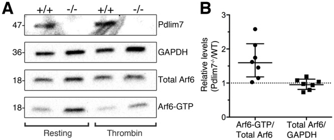
Fig 10. Loss of Pdlim7 results in unbalanced Arf6-GDP / GTP ratios.

(A) Western-blot analysis using anti-Pdlim7 antibodies of washed platelets from WT and Pdlim7-/- mice demonstrates presence of Pdlim7 proteins in WT, but not Pdlim7-/- platelet lysates (Pdlim7). Anti-Arf6 antibodies detected similar amounts of total Arf6 in Pdlim7 null platelets (Total Arf6), normalized to the internal GAPDH control (GAPDH). The protein lysate clarified supernatants were used to pull down Arf6-GTP, followed by Western bot detection using anti-Arf6 antibodies (Arf6-GTP). Comparison of resting WT to resting Pdlim7-/- platelets revealed increased Arf6-GTP to total Arf6 protein ratios following loss of Pdlim7 (A, left hand panel). (B) Graphs show individual data points normalized to control along with geometric mean and 95% confidence interval (5% CI). 95% CIs that do not cross the dotted line at y = 1 represent significant differences relative to the control at p = 0.05, n = 7. Thrombin treated platelets showed a reduced level of Arf6-GTP as compared to resting platelets as would be expected after agonist stimulation (A, right hand panel). The Western blots are representative of n = 7 (resting) and n = 2 (thrombin stimulated) independent experiments with platelets pooled from 3 mice, each experiment.
