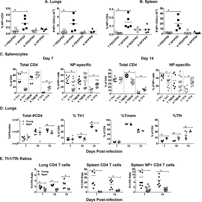
Figure 5. Age-related differences in T cells during influenza infection.
Young [Y] (2-3 mos.) and aged [A] (19-22 mos.) C57BL/6 mice were vaccinated with NP or PBS on days -31 and -21 and then infected with intranasally with 400 EID50 of PR8 influenza on day 0. On 5 dpi lungs and spleens were harvested. NP-specific CD4 T cells in the A. lungs and B. spleen were enumerated with a NP311-325/IAb MHC Class II tetramer. C. On day 0, young and aged unvaccinated mice were infected intranasally with 400 EID50 of PR8 influenza. On 7 and 14 dpi, the CD4 T cell population was further broken down phenotypically into subsets based on PSGL1 (CD162) and Ly6C expression to indicate T helper type 1 (Th1, PSGL1hi Ly6Chi), T memory (Tmem, PSGL1hi Ly6Clo) and T follicular helper (Tfh, PSGL1lo Ly6Clo) according to the scheme developed by Marshall and colleagues [38]. The percent of each subset within the total CD4 and NP-specific CD4 T cell populations is shown. D. On day 0, young and aged unvaccinated mice were infected intranasally with 400 EID50 of PR8 influenza. On 7, 10 and 12 dpi, lungs were harvested to generate single cell suspensions, which were then stained as in C.. Total CD4 numbers and percentages within Th1, Tmem and Tfh subpopulations are shown. E. Ratio of Th1/Tfh percentages from results shown in C. and D.. For all, data is representative of 2 independent experiments and each symbol represents a single animal; line shows the mean; *p < 0.05 by 2 way ANOVA with Bonferroni posthoc correction for A, B and D or Student's t test comparing young vs aged groups for C and E

