
Figure 3. Rheb activation of TOR is suppressed by RASSF1A.
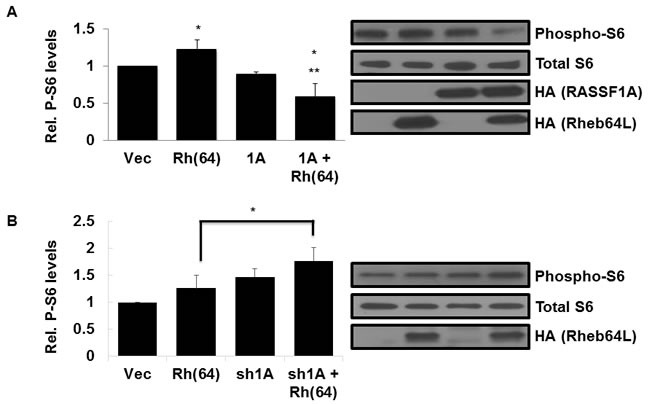
A. RASSF1A suppresses TOR activation by Rheb. (Left panel) NCI-H1299 cells described in Figure 2A were Western blotted for phosphorylated S6 expression using anti-Phospho-S6 antibodies. Immunoreactive bands were quantified by densitometry and the results plotted as a bar graph showing levels of phospho-S6 normalized to vector transfected cells. Values that are significantly different are indicated by an asterisk as follows: *, P < 0.05 compared to the value for vector control cells. **, P < 0.01 compared to the value for cells expressing Rheb(64L). (Right panel) Expression levels of each protein are shown in a Western blot. Exogenous protein expression was detected using anti-HA antibodies. The Western blot shown is representative of three independent experiments. B. Loss of RASSF1A restores S6 phosphorylation by Rheb. (Left panel) NCI-H1792 cells described in Figure 2B were Western blotted for phosphorylated S6 expression using anti-Phospho-S6 antibodies and immunoreactive bands were quantified by densitometry and the results plotted as a bar graph showing levels of phospho-S6 normalized to vector transfected cells. Values that are significantly different are indicated by an asterisk as follows: *, P < 0.05 compared to the value for cells expressing Rheb(64L). (Right panel) Expression levels of each protein are shown in a Western blot. Exogenous protein expression was detected using anti-HA antibodies. The Western blot shown is representative of three independent experiments.